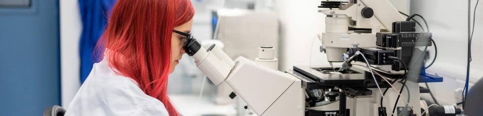


Imaging Core Facility (ICF) provides access and training to the imaging instruments in the GSH, currently including a small animal 7T MRI (Bruker), spinning disc confocal microscope (CQ1, Yokogawa), confocal microscope (SP5, Leica), fluorescent/light microscopes (AxioImager, Zeiss), light sheet microscope (UltraMicroscope Blaze, Miltenyi), in vivo imaging system (IVIS SpectrumCT, Revvity) as well as image processing stations (CQ1 off-line image analysis and CellPathFinder, Imaris Sticher and Imaris image processing software package, Living Image software +3D module).
RResponsible for the training, coordination and maintenance is Dr. Tijna Alekseeva. She serves as single point of contact for all questions related to the imaging equipment, as well as advice on the experimental set up, development of new analytical pipelines and general enquiries related to imaging. At the MRI, Dr. Alekseeva is supported by Marco Lolies who is taking care of routine measurements for internal and external users. IVIS SpectrumCT is jointly maintained with Margareta Kolaric.
All imaging instruments are primarily available to the research groups of the Georg-Speyer-Haus. However, external research groups can apply at any time.
Send your inquiry by email to t.alekseeva@georg-speyer-haus.de.
Profile
Dr. Alekseeva received her PhD in tissue engineering from University College London (United Kingdom) and worked as a postdoctoral fellow in the group of Prof. Dr. Dr. James Kirkpatrick at Mainz University Hospital and under the supervision of Prof. Fergal O’Brien at RCSI University of Medicine and Health Sciences in Dublin (Ireland). Dr. Alekseeva joined the GSH in 2017 as lab manager in the group of Dr. Sevenich. Since October 2021 Dr. Alekseeva acts as Head of the ICF.

ICF Equipment
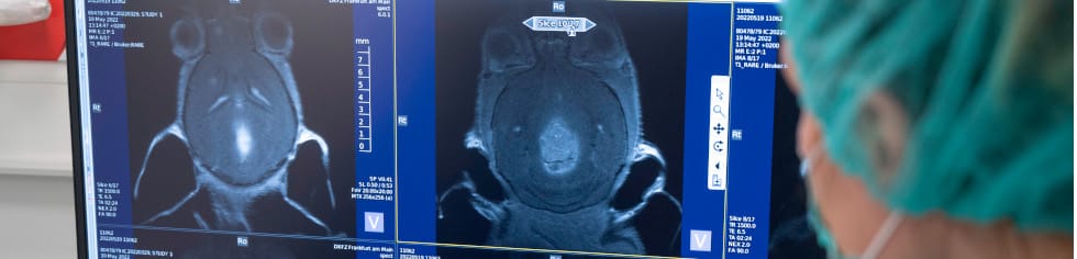
Description Small animal 7T MRI scanner
Location GSH
Book here or send email to T.Alekseeva@georg-speyer-haus.de
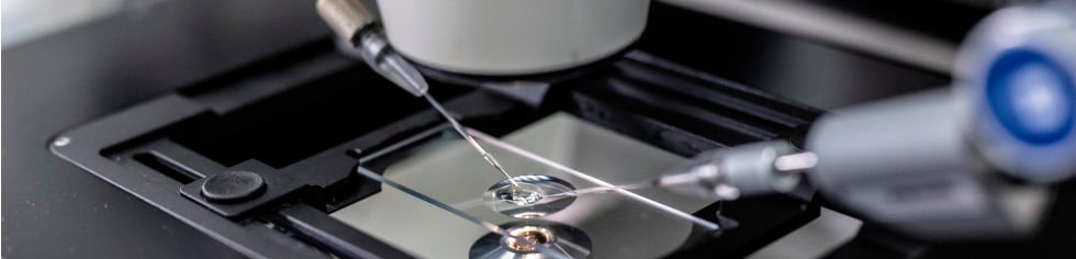
Description Confocal microscope with an inverse microscope stand
Specs 5 lasers (405 / 458, 476, 488, 496, 514 / 561 / 594 / 633), 4 objectives (10×0.4 dry; 20x/0.7 dry; 63x/1.4 oil; 63x/1.2 water)
Location GSH
Book here or send email to T.Alekseeva@georg-speyer-haus.de
Description Spinning disc confocal microscope
Specs 4 lasers (405, 488, 594,647), bright field/phase contrast, x2, x10, x20, x20 LWD, x40 and x60 objectives (all dry!), suitable for time-lapse live cell imaging, high throughput imaging
Location GSH
Book here or send email to T.Alekseeva@georg-speyer-haus.de
Description Upright dedicated fluorescent and light microscopes
Specs 4 filter cubes for 405 (DAPI), 488 (FITC), 594 (TRITC), 647 (Cy5), X1.25 (dry), x5(dry), x10(dry), x20(dry), x40(dry), x100 (oil) objectives for fluorescent and X1.25 (dry), x5(dry), x10(dry), x20(dry), x40(dry), x63 (oil) for light microscope
Location GSH
Book here or send email to T.Alekseeva@georg-speyer-haus.de
Location GSH
Send email to t.alekseeva@georg-speyer-haus.de
Description: fully automated light sheet microscope for imaging large or multiple cleared samples at subcellular resolution
Specs: White Light Laser with following filters: excitation 405/30;470/30;545/25; 560/40; 630/30;650/45; 710/75; corresponding emission: 460/40;525/50;595/40;615/40;680/30; 720/60;810/90.
Objectives: 12x (NA 0.53, WD 10 mm), 4x (NA 0.35, WD 15 mm), 1.1x (NA 0.1, WD 16 mm) + 0.6x, 1x, 1.7x, 2.5x zoom;
Location: GSH
Book here or send email to t.alekseeva@georg-speyer-haus.de.
Description: In Vivo Imaging System combining Spectral Unmixing, 2D and 3D quantitative bioluminescence and fluorescence with fast and low dose CT imaging
Specs: Click here for Specsheet.
Book here or send email to m.kolaric@georg-speyer-haus.de or t.alekseeva@georg-speyer-haus.de